Gram staining is an important and classical staining technique used in human, veterinary and ecological microbiology to differentiate bacteria into groups.
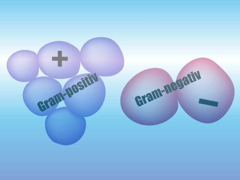
It is not easy to remember the corresponding Gram reaction due to the terms ” gram-negative” and “gram-positive”. A simple trick to remember this is to combine the words “negative” and “minus” and imagine them on a red cell background. The plus sign, on the other hand, is connected with “positive” and “plus” on a blue cell background.
How can the Gram reaction be explained? During staining, crystal violet ( = deep blue) penetrates the bacterial walls. All bacteria absorb this primary dye due to the ionic bond between the basic and acidic dye groups of the cells.
In the second staining step with iodine-iodine potassium (= Lugol solution) an iodine colour complex is formed in combination with the crystal violet.
In the third staining step, staining with alcohol, the gram positive and gram negative cells are differentiated: The iodine colour complex cannot be washed out due to the thick, rigid and narrow-pored cell walls of gram-positive bacteria. Therefore the gram-positive bacteria such as Staphylococcus aureus, Pseudomonas aeruginosa or Streptococcus pneumoniae store the deep blue dye. In contrast, in the case of the Gram-negative bacteria such as Escherichia coli, Helicobacter pylori or Spirillum volutans, the alcohol washes out the primary dye due to the coarse-meshed, thin peptidoglycan layer.
In the fourth step, the counterstain with fuchsin or saffranin, this red dye is no longer absorbed by the gram-positive bacteria and thus they appear blue under the microscope. The gram-negative bacteria, on the other hand, can absorb fuchsin or safranin and appear red under the microscope.
Whether fuchsin or saffranin is preferred as counterstain must be tested by the user himself. Some customers say that fuchsin is slightly purple and safranin, on the other hand, is clearly red. In our GRAMYfix product portfolio you will therefore find both solutions.

